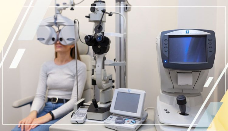
Understanding Ophthalmic Ultrasound Imaging Systems: A-Scan and B-Scan Techniques Explained
Ophthalmic ultrasound has tremendously evolved since its inception and introduction into the clinical setting in 1952. Currently, ophthalmic ultrasound imaging systems are widely used for A and B-scan contact and immersion techniques. These systems are used for intraocular characterization and orbital pathology.
It is one of the most widely used diagnostic imaging tools to diagnose multiple eye conditions. This tool also serves as a supplement to the radiology department due to its radiologic imaging functionalities of MRI and CT scanning.
In this blog, let us have a deeper look into ophthalmic ultrasound imaging systems and what is A-scan and B-scan in ocular ultrasonography.
How Does an Ophthalmic Ultrasound Imaging System Work?
The eye is a very sensitive, superficial fluid-filled structure. Hence, ultrasound is an effective and easy tool for ocular pathology and anatomy visualization. The application and principles of ultrasound in ophthalmology are similar to its application in other fields.
Sound waves with a frequency higher than 20,000 Hz (20 kHz) are created and passed through the eye. The tissues in the way reflect back the sound waves to the transducer. Once the sound waves return to the transducer, a piezo-electric crystal present in it starts vibrating. This vibration results in creating electrical impulses, which get decoded and translated into an image or other data.
A-scan and B-scan are the two major ultrasound types used in ophthalmologic practice. A-scan, also known as a time-amplitude scan, creates sound waves at 8 MHz. These sound waves are transformed into spikes, which correspond with tissue interface zones.
B-scan, also called brightness amplitude scan, creates sound waves at 10 MHz. The transducers gather the data to generate a corresponding image.
Let us now look at the A and B scans in depth.
A-Scan
Amplitude or A-scan is a technique used for ocular assessments through ultrasound. The tear film is sufficient for acoustic transmission. Hence, the need for ultrasound coupling jelly gets eliminated. The transducers send a single sound beam in this type of ophthalmology scan. Echoes that reach the transducer are transformed into a series of spikes. The height of the series of spikes is proportional to the strength of the echo.
There are various factors that determine the strength of the echo. One of them is the two tissues’ properties at an interface. The amplitude of the echo will be higher resulting in a higher spike if there are different interfaces. The amplitude of the echo will be smaller, resulting in a shorter spike, if the interfaces are similar.
The angle at which the sound wave hits the interface also impacts the spike height. If the transducer is held at a higher angle than the angle of incidence, the echoes will not return to the transducer. Hence, the height of the spike will be shorter.
Factors like the interface’s smoothness or regularity and the structure’s density through which the sound wave travels also impact how well the sound wave returns to the transducer.
More energy is absorbed by denser objects. Hence, eyes with dense cataracts block the signal’s return to the transducer. As a result, the height of the retinal spike will decrease if a cataract is present.
Healthcare professionals can use ophthalmic ultrasound imaging systems for A-scan to measure sound attenuation, spike height, regularity, and reflectivity. It will help characterize the structure of internal structures and composition based on the above-mentioned data.
B-Scan
Brightness scan or B-scan is another technique leveraged to assess ocular health through ultrasound. This scan can be executed on the anesthetized eye. Healthcare professionals can perform a B-scan on the eyelid with the use of coupling jelly while handling children or trauma cases.
Lesion’s visualization, such as shape, borders, size, and location, can be measured through a B-scan. Ophthalmic ultrasound imaging systems are used for the B-scan technique to detect foreign bodies, calcium, tumors, and retinal or choroidal detachment. The B-scan echoes are translated to dots with brightness intensity. It is proportional to the amplitude of the echo. B-scan is essential for imaging the tumors of the anterior orbit, myositis, and other ophthalmic veins.
Just like the A-scan, the B-scan has a good sensitivity but poor resolution. Healthcare providers can get the best image quality if the lesions are centered in the image.
To Summarize
Ophthalmic ultrasound imaging systems are one of the most widely used ocular ultrasonography tools for imaging. It helps get the globe length to measure the corrective intraocular lens power needs. Additionally, it is also helpful to calculate tumor measurements, such as choroidal melanomas, imaging of lens dislocation, and determining retinal detachment.
Ocular ultrasonography is beneficial in cases where the fundus is blocked from visualization, especially in patients with dense cataracts. Using ophthalmic ultrasound imaging systems will help to detect ocular melanoma in its early stages.
One of the biggest advantages of ophthalmic ultrasound is enhanced visualization of structures blocked by opaque substances like dense cataracts or vitreous hemorrhage. Additionally, it helps healthcare providers have real-time information about conditions like retinal detachment. Ophthalmic ultrasound is affordable, easily accessible, safe, and does not expose patients to radiation.


